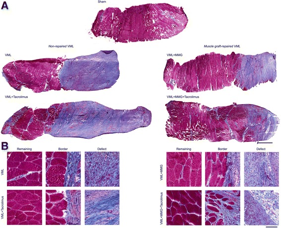
Fig. 1.

Representative histologic micrographs of Masson’s Trichrome stained (connective tissue is blue; nuclei are purple; skeletal muscle fibers are red) porcine peroneous tertius (PT) muscle following VML injury and repair. While all PT muscles indicate gross fibrosis following VML injury, only the muscle graft-repaired displayed areas of likely regenerated fibers. There were no apparent differences due to treatment with tacrolimus. (a) Each sample represents a full thickness sample through the muscle. Scale bar is 2 mm; all images are at the same magnification. (b) Representative inserts from the remaining muscle, border, and defect area of the samples are displayed. Scale bar is 100 μm; all images are at the same magnification
