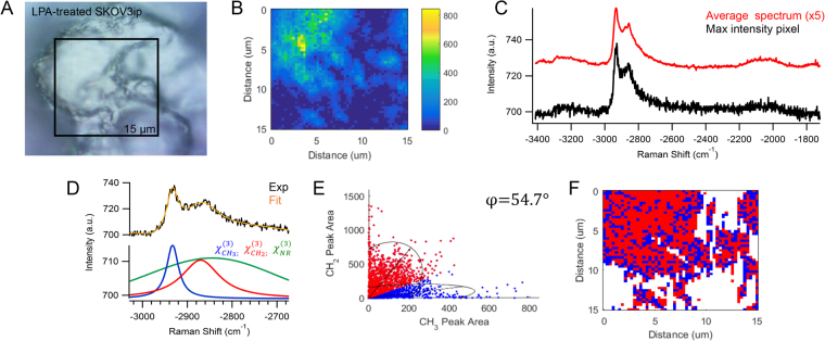
Figure 8.
(A) Brightfield image of LPA-treated SKOV3ip cells on the surface of the MCA is consistent with (B) the CARS image of −2930 cm−1 peak area. (C) The maximum intensity CARS spectrum and average spectrum of the map show varying contributions of CH2 and CH3 signal. (D) An example of the nonlinear peak fit (orange) of the CARS intensity (black) has contributions from the nonresonant background (green) and resonant vibrational bands at −2850 cm−1 (red) and −2930 cm−1 (blue). (E) The frequency scatter plot exhibits overlapping spectral clusters with 95% confidence ellipses and angle, φ, corresponding to the spectral separation of lipids (red) and proteins (blue). (F) The reconstructed map shows the chemical distribution of the segmented lipid (red) and protein (blue) pixels. Experimental parameters: Pump = 720 nm, 25.20 mW; Supercontinuum = 785–945 nm, 3.80 mW; Acquisition time = 500 ms/pixel; Map size = 51 × 51 pixels; Steps = 300 nm; Objective = 100x, 0.9 NA.

