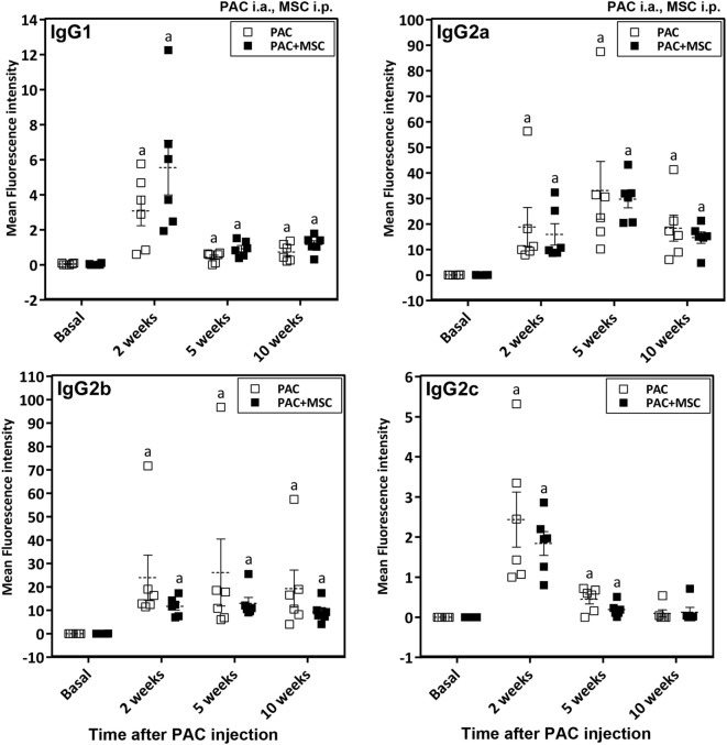
Figure 5.
Changes in IgG antibody subtypes in Lewis rats injected intraarticularly (i.a.) with porcine articular chondrocytes (PAC) only or posttreated with mesenchymal stem cells (MSC) intraperitoneally (i.p.). The study included samples from the PAC and PAC + MSC cohorts collected at baseline, 2, 5, and 10 weeks after PAC injection (Figure S1C in Supplementary Material). In particular, the PAC cohort received only PAC i.a., whereas the PAC + MSC rats were injected with MSC i.p. 3 weeks after PAC i.a. injection. Anti-PAC IgG1, IgG2a, IgG2b, and IgG2c antibody reactivity was determined by flow cytometric analysis in sera of all experimental rats at 1%. The mean FL-1 fluorescence intensity after subtracting the background (reactivity of secondary antibody alone) is shown as mean ± SEM for the PAC and PAC + MSC cohorts (n = 6). Statistically significant differences were observed using the Wilcoxon test for the assessed time points and subtypes relative to corresponding baseline levels as indicated (ap ≤ 0.05). No significant differences were observed using the Mann–Whitney U test when comparing results from the PAC and PAC + MSC cohorts.

