Figure 3.
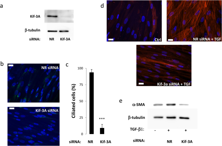
(a) aAPs were treated with a non-relevant (NR) or a Kif-3A siRNA as described in Material and Methods. (a) Kif-3A expression was analyzed by Western blot. (b) Cells were fixed and Ac-Tub (in green) was revealed by immunocytochemistry. The white bar represents 20 μm. (c) Percentages of ciliated cells were measured (n = 3 ***p < 0.001). Cells were treated or not with TGF-β1 for 3 days and α-SMA expression was analyzed by (d) immunofluorescence or (e) Western blot.

