Figure 1.
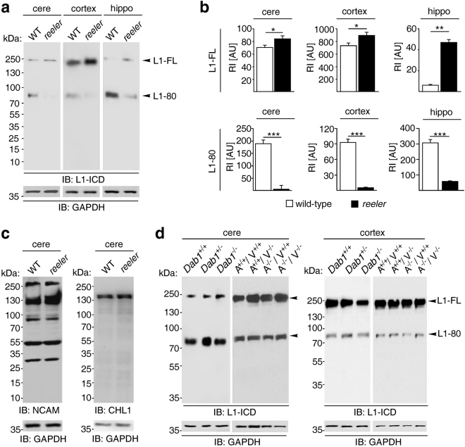
L1-80 levels are decreased in reeler mice. (a) Immunoblot analysis of homogenates from cerebellum (cere), cerebral cortex (cortex) and hippocampus (hippo) of 6-day-old wild-type (WT) and reeler mice with an antibody against the intracellular L1domain (L1-ICD). L1-FL: full-length L1. (b) Quantification of L1-FL and L1-80 levels in homogenates from cerebellum, cerebral cortex and hippocampus of wild-type and reeler mice. Mean values + SEM from 6 independent experiments and differences between groups are shown (*p < 0.05, **p < 0.01, ***p < 0.005; two-tailed t-test). RI: relative intensity in arbitrary units (AU). (c) Unaltered NCAM and CHL1 expression levels in cerebellar homogenates from wild-type (WT) and reeler mice. (d) Unaltered L1-80 levels in cerebellar and cerebral cortex homogenates from wild-type (Dab1 +/+), heterozygous (Dab1 +/−), and Dab1 knock-out (Dab1 −/−) mice, and in homogenates from ApoER2-VLDLR double knock-out (A−/−V−/−), heterozygous (A+/+V−/−, A−/−V+/+) and wild-type (A+/+V+/+) littermates. (a,c,d) Representative immunoblots out of 6 independent experiments are shown and display all L1, CHL1 and NCAM forms. GAPDH antibody was used to control loading and only the regions of the blots with GAPDH bands are shown.

