Figure 2.
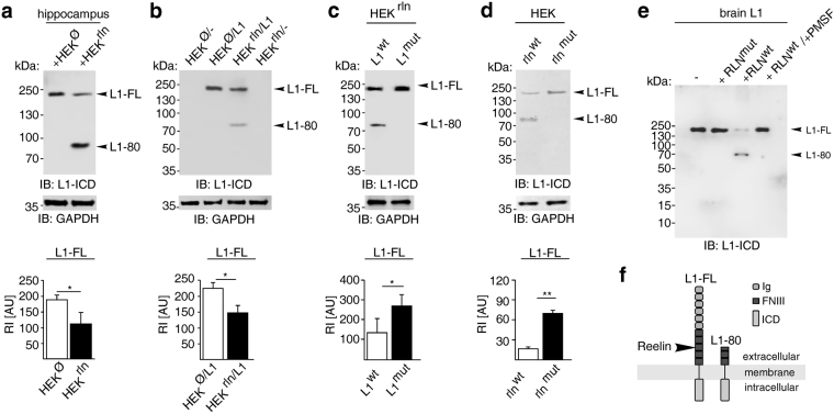
Reelin cleaves L1. (a) Freshly homogenized reeler hippocampus was treated with supernatants from HEK cells expressing (HEKrln) or lacking (HEKØ) Reelin. (b) HEKrln and HEKØ cells were transfected with wild-type L1 (L1) or mock DNA (−). (c) HEKrln cells were transfected with wild-type (L1wt) or mutated (L1mut) L1. (d) HEK cells were co-transfected with wild-type L1 and wild-type Reelin (rlnwt) or mutated Reelin (rlnmut). (a–d) Representative immunoblots of cell lysates out of six independent experiments using an antibody against the intracellular L1 domain (L1-ICD) and the GAPDH antibody are shown. The cropped blots display all L1 forms or the GAPDH band. Mean values + SEM from 6 independent experiments are shown for the levels of full-length L1 (L1-FL) and differences between groups are shown (*p < 0.05, **p < 0.0001; One-way ANOVA with Tukey’s Multiple Comparison Test). RI: relative intensity in arbitrary units (AU). (e) Purified brain L1 was incubated with heparin-purified wild-type (RLNwt) or mutated (RLNmut) Reelin protein in the absence or presence of the serine protease inhibitor PMSF (+PMSF) and then subjected to immunoblot analysis with L1 antibody recognising the intracellular domain (L1-ICD). (f) Schematic representation of L1 structure and cleavage by Reelin. Shown are the extracellular Ig and FN III domains, the transmembrane and intracellular domain (ICD).

