Figure 1.
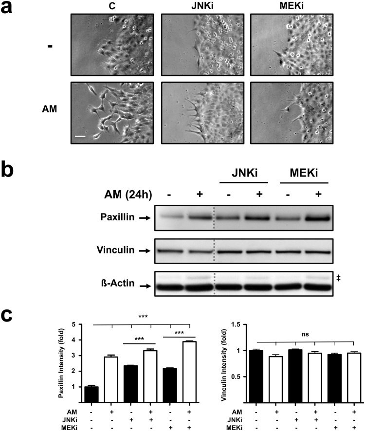
Amniotic membrane (AM) promotes cell protrusion generation and Paxillin expression in migrating Mv1Lu cells. (a) Detailed pictures of the migrating edge of artificial wound assays treated with AM in combination with inhibitors. Scale Bar 50 µm. (b) Western Blot of total protein extracts from sub-confluent Mv1Lu cells cultured in the presence of AM and/or inhibitors and collected after 24 hours. The dashed grey lines indicate that two distant parts of the very same blot were put together. ß-actin was used as loading control. (‡) Unspecific bands. (c) Relative protein level plots generated from Western Blot quantification. C: serum starvation; JNKi: SP600125; MEKi: PD98059. Asterisks denote statistically significant differences between conditions according to ANOVA statistical analysis: (***) p < 0.001; (ns) not significant. All experiments were repeated at least three times. Representative pictures and results are shown.

