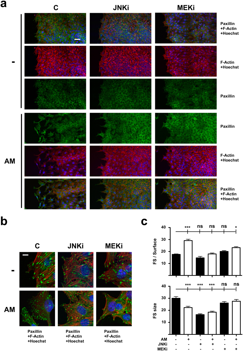
Figure 2.
Amniotic membrane (AM) treatment alters Paxillin distribution and focal structures arrangement in migrating Mv1Lu cells. (a) Confocal microscopy images of Mv1Lu cells at the migrating edge of artificial wound assays. Scale Bar 50 µm. Juxtaposed field images were taken to show migration front from a wider perspective. (b) 2.5x magnified detail from merged pictures in (a). Scale Bar 10 µm. (c) Plots for average number and size of focal structures detected at the migrating leading edge. Asterisks denote statistically significant differences between conditions according to ANOVA statistical analysis: (*) p < 0.05, (**) p < 0.005 and (***) p < 0.001; (ns) not significant. Paxillin: green; Actin: red; Nuclei: blue; C: serum starvation; JNKi: SP600125; MEKi: PD98059; FS: focal structures. All the experiments were repeated at least three times. Representative pictures are shown.

