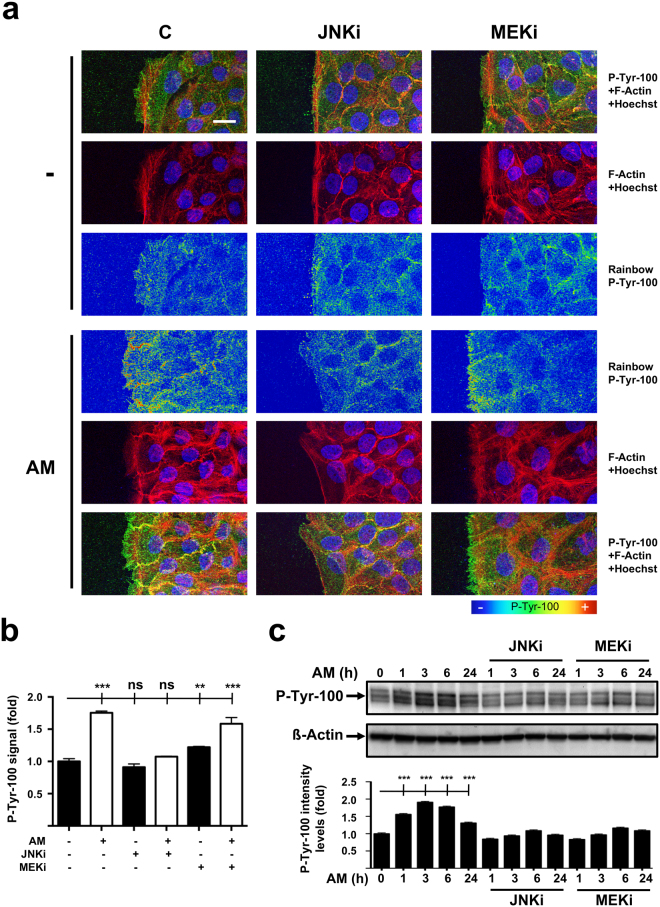
Figure 5.
Amniotic membrane (AM) promotes FAK activation and detection at focal structures in migrating HaCaT cells. (a) HaCaT cells at the migrating edge of artificial wound assays show increased p-tyr-100 immune-staining when AM was present. P-tyr-100 immune-staining signal was converted into pseudo-color rainbow scale to display fluorescence-intensity variations. Fluorescent signal was converted using Rainbow feature from ZEN software, on a linear mode and covering the full range of the data. Co-staining with phalloidin and Hoechst-33258 was used to show the cell structure and nuclei, respectively. P-tyr-100: pseudo-color; p-tyr-100: green; Actin: red; Nuclei: blue. Scale Bar 25 µm. (b) Relative fluorescence level plot generated from average p-tyr-100 signal intensity data obtained at the migrating leading edge. (c) Western Blot of total protein extracts from sub-confluent HaCaT cells cultured in the presence of AM and/or different inhibitors collected for a 24 hours time course. ß-actin was used a loading control. Relative protein level plots were generated from Western Blot quantification data. C: serum starvation; JNKi: SP600125; MEKi: PD98059. Asterisks denote statistically significant differences between conditions according to ANOVA statistical analysis: (***) p < 0.001; (ns) not significant. All the experiments were repeated at least three times. Representative pictures and results are shown.

