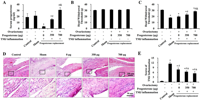
Figure 1.
Progesterone attenuated CFA-induced TMJ mechanical hyperalgesia and inflammation. (A) Serum levels of progesterone in intact rats and progesterone-treated ovariectomized rats. Among the ovariectomized rats, serum levels of progesterone were lowest in 0 μg-P4 treated rats and increased in a dose-dependent manner with the increasing of injected progesterone. *P < 0.05 versus control and sham; # P < 0.05 versus 0 μg; & P < 0.05 versus 20 μg, by two-way analysis of variance (ANOVA). (B,C) Head withdrawal threshold before (B) and after (C) TMJ inflammation. Progesterone ameliorated the TMJ inflammation–induced decrease in the head withdrawal threshold. (D) Representative images of the TMJ regions in female rats. Small and large black frames indicate the original and the magnified areas, respectively. Following induction of inflammation for 24 hours, TMJs of the sham-OVX and the 0-μg-P4 group showed typical features of synovitis. Compared with the 0-μg-P4 group, the proliferation of synoviocytes was less than that in 350-μg-P4 group. In 700-μg-P4 group, the synovial lining layer was thin and infiltration of mononuclear cells around the synovial membrane was not evident. (E) Progesterone attenuated CFA-induced TMJ inflammation. Scores for TMJ inflammation decreased with increasing doses of P4. *P < 0.05 versus control; # P < 0.05 versus sham; & P < 0.05 versus 0 μg, by two-way ANOVA.

