Figure 5.
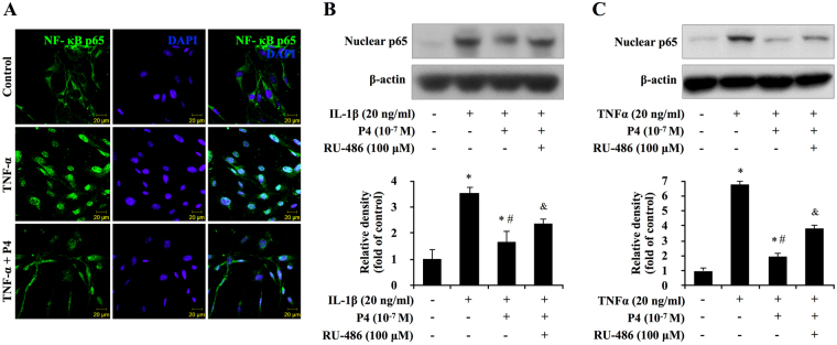
Blocking progesterone receptor on NF-κB p65 nuclear translocation in primary cultured synoviocytes. (A) Immunofluorescence of p65 in TNF-α and P4 treated synoviocytes. The subcellular translocation of p65 was examined using confocal microscopy 24 hours after the induction of TNF-α in synoviocytes. Fluorescence signal of p65 (green) was observed in the cytoplasm of the synoviocytes in control group. In contrast, the fluorescence signal of p65 translocated to nucleus in TNF-α-induced group. The green staining of NF-B p65 changed into watery blue after merging with the blue staining of the nucleus, counterstained with 4,6-diamidino-2- phenylindole (DAPI; blue). In TNF-α and P4 treated group, translocation activity was partially blocked by pretreatment of P4 In TNF-α and P4 treated group, with green staining not only observed in nucleus but also in cytoplasm of synoviocytes. (B,C) Blocking progesterone receptor partially reversed TNF-α/IL-1β-induced transcription of cytokines in primary synoviocytes. The synoviocytes were treated with TNF-α/IL-1β and P4 for 24 h after pretreated with progesterone receptor antagonist RU-486. The expression of p65 was evaluated by western blot or real-time PCR. The middle panels show the quantification of p65 protein presented as the relative density compared with the control group. β-actin was served as an internal control for equal loading. *P < 0.05 versus control; # P < 0.05 versus the group treated with TNF-α/IL-1β; & P < 0.05 versus other groups.

