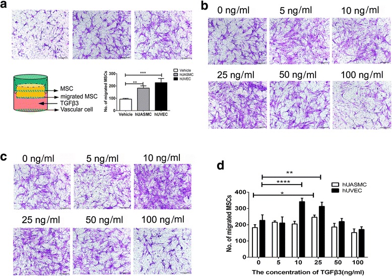
Fig. 2.

Transwell assay for hBMSC migration. a Analysis for the migration of hBMSCs with or without hUVECs and hUASMCs. b In the coculture system of hBMSCs and hUASMCs, representative light photomicrographs of migrated hBMSC induced by 0-100 ng/ml TGFβ3 after 24-hour incubation. c In the coculture system of hBMSCs and hUVECs, representative light photomicrographs of migrated hBMSCs induced by 0-100 ng/ml TGFβ3 after 24-hour incubation. d Quantitative analysis of migrated cell density for (c) and (d). Migrated cells were stained purple with crystal violet. Scale bar: 100 μm. *P < 0.05, **P < 0.01, ***P < 0.005, ***P < 0.001. hUASMC human umbilical artery smooth muscle cell, hUVEC human umbilical vein endothelial cell, MSC mesenchymal stem cell, TGFβ3 transforming growth factor beta-3
