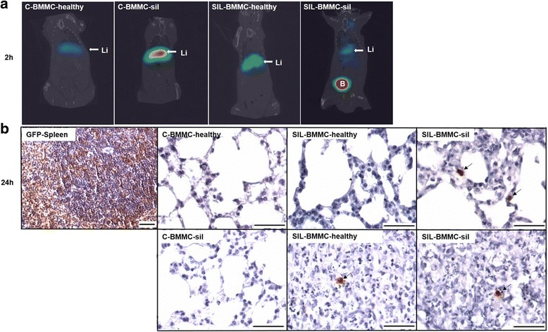
Fig. 2.

Biodistribution and homing of bone marrow mononuclear cells (BMMCs). a Representative coronal whole-body SPECT/CT images of control (C) and silicotic (SIL) animals 2 h after intravenous administration of 99mTc-BMMCs derived from heathy or silicotic (sil) donors. 99mTc-BMMCs migrate to the liver in all experimental groups (Li). B bladder. n = 4 animals per group. b Representative photomicrographs of lung parenchyma, granuloma, and spleen after immunohistochemistry with green fluorescent protein (GFP) antibody. Rare inflammatory GFP+ cells (black arrows) were observed in lung parenchyma and in granulomas of the SIL-BMMC groups 24 h after treatment with BMMCs from GFP+ mice. Spleens of GFP male mice were used as positive control. Bars, 100 μm. n = 4 animals per group
