Fig. 1.

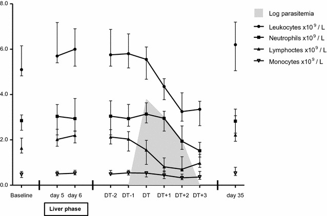
Changes in total and differential leukocyte counts in the 20 subjects who developed malaria in CHMI-b. The data are shown as medians (dots) and interquartile ranges (whiskers). The data from DT − 2 until DT + 3 were synchronized on DT

Changes in total and differential leukocyte counts in the 20 subjects who developed malaria in CHMI-b. The data are shown as medians (dots) and interquartile ranges (whiskers). The data from DT − 2 until DT + 3 were synchronized on DT