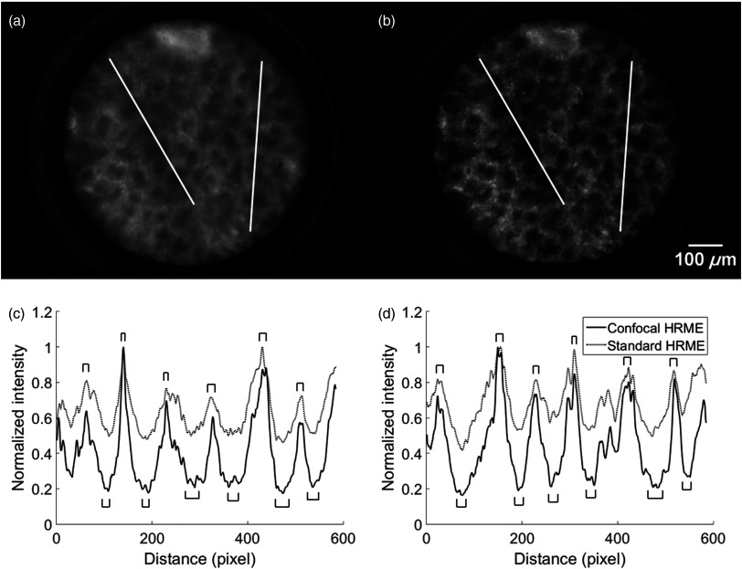
Fig. 6.
Ex vivo images of mouse columnar epithelium using (a) the standard and (b) confocal HRME. Profiles are shown for the line scans on the (c) left and (d) right. The brackets indicate the glandular walls and lumens. The resulting gland-to-lumen ratio was higher in the confocal than the standard HRME ( in standard and in confocal; ).

