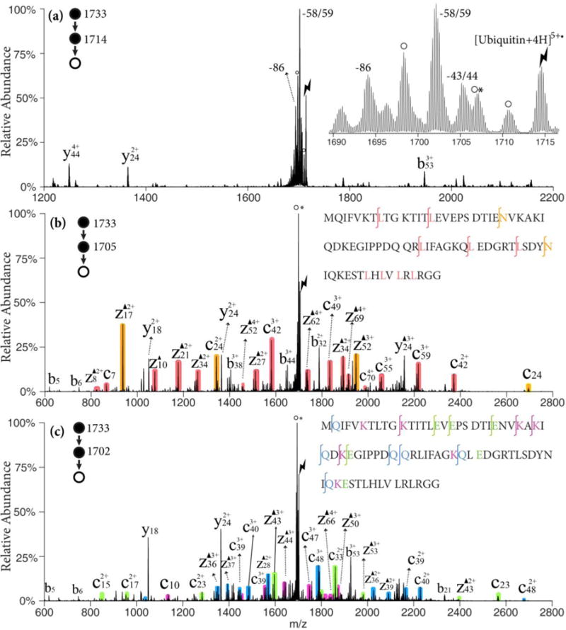
Figure 3.

(a) CID of [ubiquitin +4H]5+• ion derived from ion/ion reaction between [ubiquitin +6H]6+ and SO4−•; Insert: zoom-in of the radical side chain loss region. (b) CID of the 43/44 Da loss from [ubiquitin +4H]5+•; (c) CID of the 58/59 Da loss from [ubiquitin +4H]5+•; Black triangles indicate fragment ions containing a Dha residue. Degree signs indicate water losses whereas asterisks indicate ammonia losses. Red, orange shaded peaks indicate c- and z-fragments N-terminal to leucine and asparagine. Violet, green and blue shaded peaks indicate c- and z-fragments N-terminal to lysine, glutamine and glutamic acid residues, respectively. Lightning bolts indicate species subjected to activation.
