
Figure 2. Combined injury results in overgrowth of Enterobacteriaceae despite increased epithelial cell AMPs.
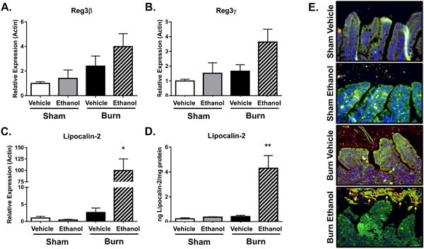
Real-time PCR analysis of AMP transcripts for Reg3β, Reg3γ, and lipocalin-2 following the combined injury, sham (A-C). Lipocalin-2 ELISA from total small intestine tissue (D), n = 3-11 animals per group. Specific fluorescent probes were used to perform FISH staining on total bacteria (red labeled probe) and Enterobacteriaceae (green labeled probe) on sections from distal ileum (E). Enterobacteriaceae appear yellow (arrows) due to the overlap of total bacteria (red) and Enterobacteriaceae (green) probes. FISH staining from one representative experiment, n = 3-6, *p < 0.05, **p < 0.01, by One-Way ANOVA with Tukey post-hoc compared to all groups.