Figure 2.
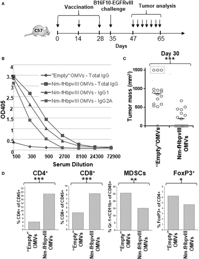
Immunogenicity and protective activity of Nm-fHbpvIII-outer membrane vesicles (OMVs). (A) Schematic representation of immunization and challenge schedules in C57BL/6 mice. (B) Anti-EGFRvIIIpep antibody titers in C57BL/6 mice immunized with “Empty” OMVs and with Nm-fHbpvIII-OMVs. Sera from mice immunized as reported in (A) were pooled and total IgGs, IgG1, and IgG2a were measured by ELISA, coating the plates with synthetic EGFRvIIIpep (0.5 μg/well). (C) Analysis of tumor development in C57BL/6 mice immunized with “Empty” OMVs and with Nm-fHbpvIII-OMVs. The figure reports the tumor size in each mouse as measured at day 30 after challenge with 0.5 × 10⁵ B16F10EGFRvIII cells. *** indicates a statistically significant difference of P < 0.001. (D) Analysis of tumor-infiltrating cell populations. At the end of the challenge experiment, two tumors/group were randomly selected and the percentage of infiltrating CD4+ T cells, CD8+ T cells, MDSCs, and Tregs was determined by flow cytometry, as described in Section “Materials and Methods” (*P < 0.05; **P < 0.01; ***P < 0.001).

