
FIGURE 4.
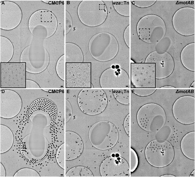
Outer membrane vesicle arrangement and distribution in Vibrio vulnificus. (A,D) A 3 × 3 image montage of wild-type CMCP6 showing characteristic net-like OMV arrangement around the cell with regular spacing between OMVs and a pronounced empty zone proximal to the outer membrane. Inset in (A) is magnified 2.5×. Black dots in (D) represent centers of OMVs seen unobstructed in (A). (B,E) A 3 × 3 image montage of the wza::TnPhoA unencapsulated (FLA1009) mutant showing the more random distribution of OMVs around the cell. Inset in (B) is magnified 5×. (C,F) A 3 × 3 image montage of ΔmotAB flagellar motor mutant (FLA674) showing a similar OMV arrangement and distribution as wild-type, but having reduced OMV numbers. Inset in (C) is magnified 2.5×. Image montage area = 7.5 μm2. Scale bars, 200 nm.