
FIGURE 7.
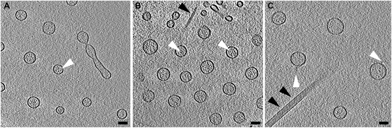
Contents of Vibrio vulnificus OMVs. (A–C) Averaged central tomographic slices of OMVs. All show electron dense contents inside the vesicle, as well as occasional very small densities on the outside of the membranes, white arrowheads. (A) Contains a dividing tube. Single black arrowhead in (B) indicates the pilus. Double black arrowheads in (C) indicate the flagellum. Scale bars, 50 nm.