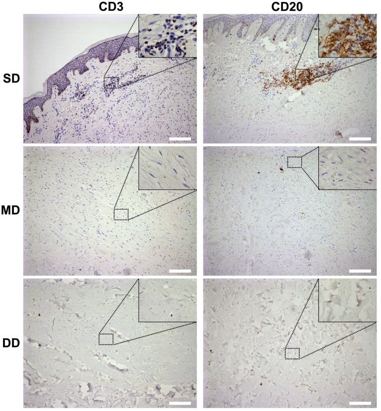
Figure 2.
Lymphocyte infiltration in the keloid dermis at different depths. CD3+ T-lymphocytes and CD20+ B-lymphocytes in keloid was detected by immunohistochemistry staining. SD, the superficial dermis of keloids; MD, the middle dermis of keloids; DD, the deep dermis of keloids. Scale bar = 200 μm.

