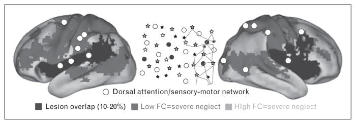
FIGURE 1.
Patterns of resting state functional connectivity associated to spatial neglect at acute stage. The white circles indicate the regions of interest (ROIs) belonging to dorsal attention/sensory motor networks, whose functional connectivity is associated to the degree of the rightward spatial biases in attention typical of neglect patients (schematically illustrated in central inset, in a large sample of stroke patients (n = 84). Spatial neglect is associated with two abnormal patterns of functional connectivity derived from the white ROIs: reduced interhemispheric functional connectivity with regions of dorsal attention/sensory motor networks in the opposite hemisphere (blue voxels); increased functional connectivity (reduced segregation) with regions of default mode/fronto-parietal networks (orange voxels). Note that dorsal attention/sensory motor and default mode/fronto-parietal networks are typically anticorrelated in healthy individuals, hence, the increased functional connectivity should be read as reduction of ‘segregation’ between two sets of networks. Black color indicates voxels damaged in 10–20% of patients. Reproduced with permission [15].

