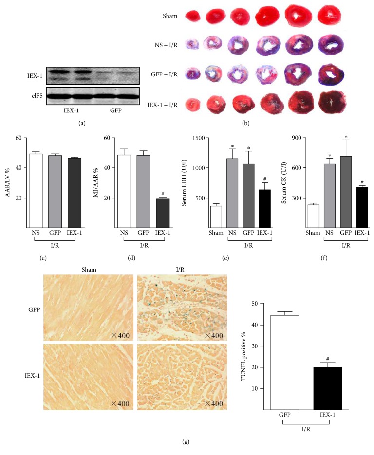
Figure 3.
IEX-1 overexpression attenuates myocardial infarction. Rats were injected with saline (NS) (n = 7), Ad-GFP (n = 11), or Ad-IEX-1 (n = 8) for 4 days, then underwent I/R (30 min/24 h). (a) Western blot analysis confirmed overexpression of IEX-1 in the left ventricle after four days of local adenovirus delivery. (b) Representative images of the infarct heart stained by Evans blue and TTC. (c) Ratios of AAR to LV and (d) MI to AAR were shown. (e) Serum LDH and (f) CK activity were measured at the end of 24-hour reperfusion. (g) After subjected to I/R, hearts were excised for slicing, then TUNEL staining was performed for assessing cardiac cell apoptosis post-I/R. ∗P < 0.05 versus Sham, #P < 0.05 versus GFP.

