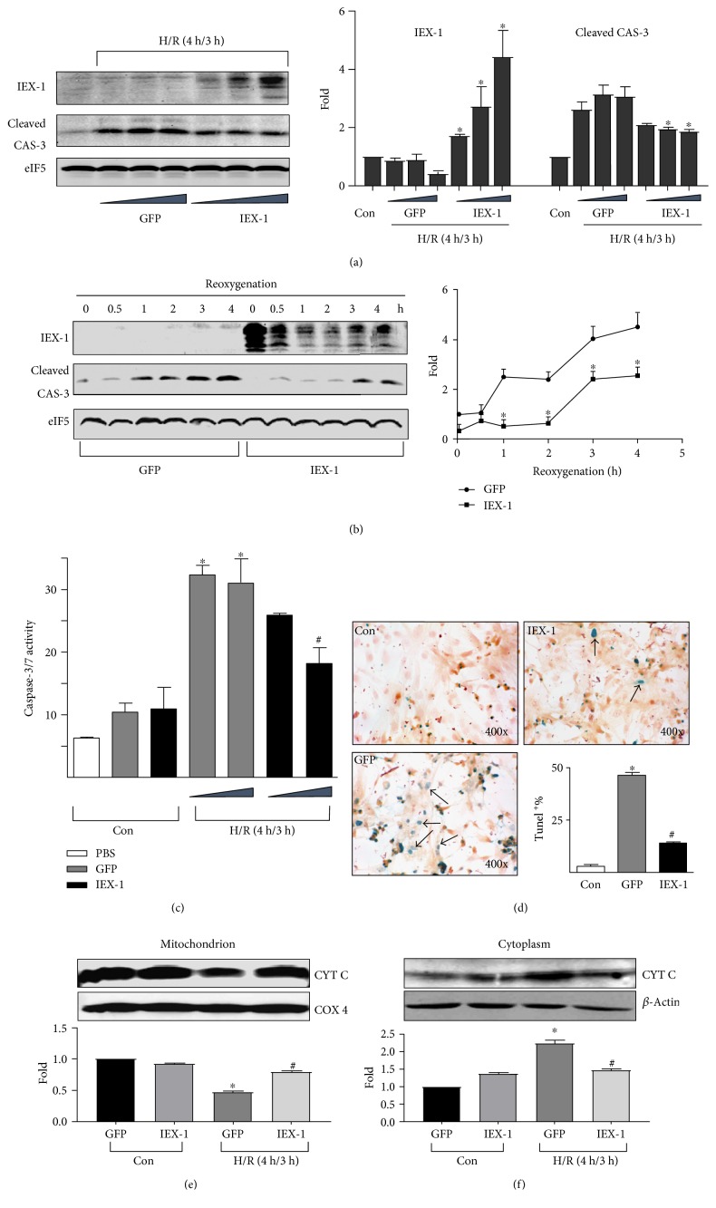
Figure 7.
IEX-1 overexpression attenuated H/R-induced cardiomyocyte injury. (a) Cardiomyocytes were infected with Ad-GFP or Ad-IEX-1 (5, 10, and 20 MOI), then underwent H/R. Results are from one representative experiment of three. (b) Cardiomyocytes were infected with 10 MOI adenoviruses, then underwent hypoxia (4 h) and reoxygenation for the indicated time. (c) Cardiomyocytes together with the medium were collected after H/R, total caspase-3/7 activity was detected. Triangle represents adenovirus infection of 5 and 10 MOI. (d) Cardiomyocytes underwent H/R, and TUNEL staining was performed to assess cell apoptosis. Arrows show TUNEL-positive cells, and the ratio of TUNEL-positive cells was calculated. Results are from one representative experiment of four. (e) Mitochondrial and (f) cytoplasmic fractions were isolated from cardiomyocytes, and proteins were probed with an anti-cytochrome c antibody. eIF5, COX4, or β-actin expression was a control for protein loading. Each column represents results from at least 3 independent experiments. ∗P < 0.05 versus control (Con or corresponding time points), #P < 0.05 versus the corresponding GFP group.

