Figure 9.
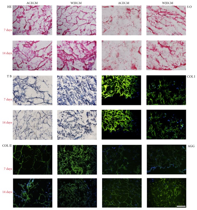
Histological and immunofluorescent staining of chondrocytes cultured on WJECM and ACECM scaffolds 7 and 14 days after seeding (scale bar = 200 μm). H&E staining showed that the cells were distributed evenly and secreted ECM in both of the scaffolds. Safranin O and toluidine blue staining showed that chondrocytes in the WJECM scaffold showed more intense staining than those in the ACECM scaffold at 7 or 14 days. At 7 and 14 days, both scaffolds were positive for collagen I, collagen II, and aggrecan.

