
Figure 3. Effects of uPAR mediated fibroblast targeting on fibroblast-tumor cell interactions in vitro.
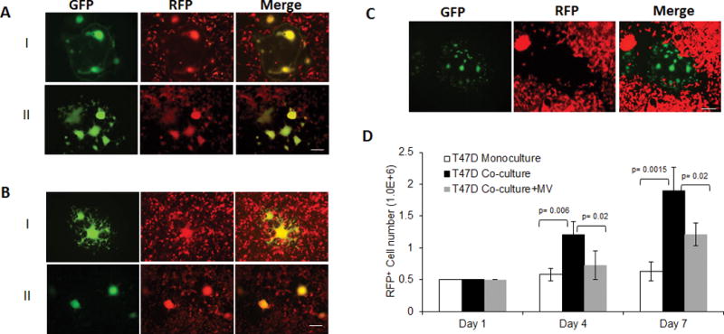
CAF19 (A) were infected with MV-h-uPA (MOI=1) as in methods, and then overlaid on RFP-expressing MDA-MB-231 (A, I) and HT-29 (A, II) cancer cells. Mixed (double-color) syncytia were demonstrated at 48h incubation by fluorescent microscopy. (B) In a similar way, MV-m-uPA infected 3T3 cells were overlaid on RFP-expressing murine 4T1 (B, I) or CT-26 (B, II) cells and yellow syncytia were demonstrated at 48h incubation by fluorescent microscopy. Scale bar = 100 μm. (C). MV-m-uPA infected murine 3T3 cells were overlaid onto human T47D, RFP expressing T47D breast cancer cells. Note that no yellow syncitia was observed in the merged image. Scale bar = 100 μm. (D) Effects of murine fibroblast specific viral targeting on human T47D cancer cell growth in vitro using an in vitro 3-D collagen co-culture model (see methods). Bars represent averages +/− SD of triplicate experiments.