
Figure 4.
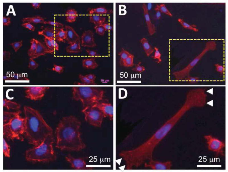

Morphology of keratinocytes on PCL/collagen nanofibrous matrices alone or with collagen gel coating. A–H) Immunofluorescent staining of intracellular cytoskeleton protein of F-actin (Red) and nuclei with DAPI (purple/blue) (A, C: PCL/collagen nanofiber alone; B, D: with collagen gel coating), and focal adhesion proteins of vinculin (Green) (E, G: PCL/collagen nanofiber alone; F, H: with collagen gel coating). Arrowhead: keratinocyte lamellipodium; arrow: spatial distribution of vinculin. I) Percentage of polarized keratinocytes (n=3) ** Statistically significant, p < 0.001