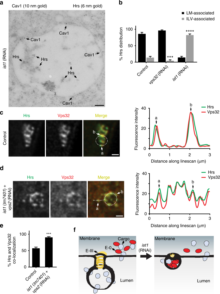
Fig. 6.
Ist1 inhibits the incorporation of ESCRT-0 into ILVs. a Immunogold labeling was used to determine the distributions of Cav1 (10 nm gold) and Hrs (6 nm gold) in one-cell stage embryos depleted of Ist1 following high-pressure freezing. The image shown is representative of 64 MVEs analyzed. Bar, 100 nm. b Quantification of the distribution of Hrs at the limiting membrane and within MVEs in control (n = 31), Vps32-depleted (n = 36), and Ist1-depleted (n = 27) one-cell stage embryos. Standard error of the mean is reported in each case. ***p < 0.0005; ****p < 0.0001 compared to control using a two-way ANOVA with Dunnett’s test. c, d Control (n = 104) and ist1 (tm7401) mutant embryos (n = 111) expressing GFP::Cav1 and depleted of Vps2 using RNAi were fixed and stained using antibodies directed against Hrs and Vps32. Samples were imaged using STED microscopy, and representative images are shown. Linescan analysis was conducted to measure the relative distributions of Hrs and Vps32 at endosomes (n = 76 linescans per condition). Specific sites along each linescan are highlighted. Bars, 500 nm. e Quantification of co-localized Hrs and Vps32 under conditions described in panel d. Standard error of the mean is reported in each case. ***p < 0.001 using a t-test. f A model of MVE biogenesis in the presence and absence of Ist1. Under normal conditions, cargoes are sorted into membrane subdomains that are internalized via ESCRT-III activity. However, following depletion of Ist1, cargo retention within ESCRT-III subdomains is impaired and ESCRT-0 becomes engulfed into ILVs of reduced size

