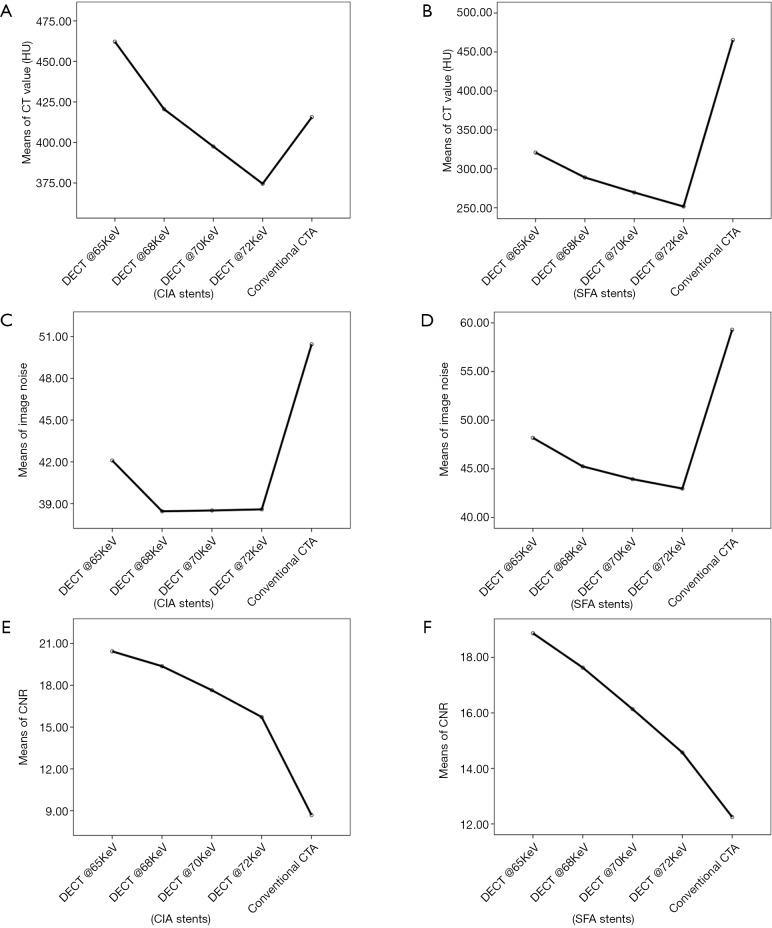
Figure 2.
A line graph of comparison between the 4 VMS and conventional CTA for stents at CIA and SFA. (A,B) CT attenuation (HU) shows the highest CT value at the lower keV; (C,D) image noise demonstrates that the lowest image noise was found at 72 keV protocol and the highest image noise at the conventional CTA, followed by 65 keV protocol; and (E,F) contrast-to-noise ratio (CNR) of CIA shows that CNR decreased with increase in VMS, however, all of the VMS were found to be better than conventional CTA. VMS, virtual monochromatic spectral; CTA, CT angiography; CIA, common iliac artery.

