Figure 4.
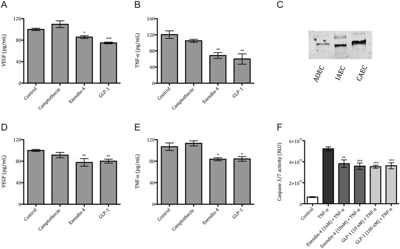
VEGF, TNF-α concentration in ovarian cancer cells and Caspase 3/7 activation in endothelium. VEGF protein levels in SKOV-3 (A) and CAOV-3 (D) and TNF-α protein levels in SKOV-3 (B) and CAOV-3 (E) after 24 h incubation with Camptothecin (100 nM), Exendin-4 (50 nM) and GLP-1 (100 nM). Representative Western blot image of GLP-1R expression in human Aortic Endothelial Cells (AoEC), Iliac Artery Endothelial Cells (IAEC) and Coronary Artery Endothelial Cells (CAEC) (C). Caspase 3/7 activation in TNF-α (10 ng/mL) stimulated IAEC after incubation with Exendin-4 (1 nM and 10 nM), and GLP-1 (10 nM and 100 nM) (F). Mean values ± s.e.m. are shown. n = 3–6 per group. *P < 0.05, **P < 0.01, ***P < 0.001 TNF-α vs different conditions. One-way ANOVA followed with Dunnett’s post hoc.

 This work is licensed under a