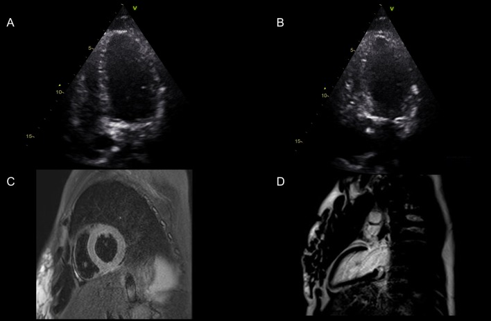
Figure 2.
Images (A) and (B) display the apical 4 chamber view in end diastole and end systole The basal anteroseptal, basal inferolateral and basal inferior segments had reasonable contractility, and the mid-inferior segment was hypokinetic. The remaining segments were severely hypokinetic/akinetic. Visually estimated LVEF = 10–15%. (C and D) Cardiac MRI showed improvement in LV systolic function with no myocardial oedema on T2-weighted sequences (Fig. 2C), and no late gadolinium enhancement to suggest myocardial fibrosis (Fig. 2D).

 This work is licensed under a