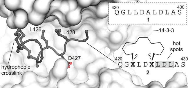
Figure 1.

Sequence of linear peptide 1 and crystal structure of cyclic peptide 2 (dark gray) bound to 14–3–3ζ (light gray, PDB ID 4n84). Cross-link and hotspot residues (L426, D427, and L428) are shown explicitly. Peptide sequence of 2 and chemical structure of cross-link are shown (Residues are numbered in accordance to PDB ID 4n84).
