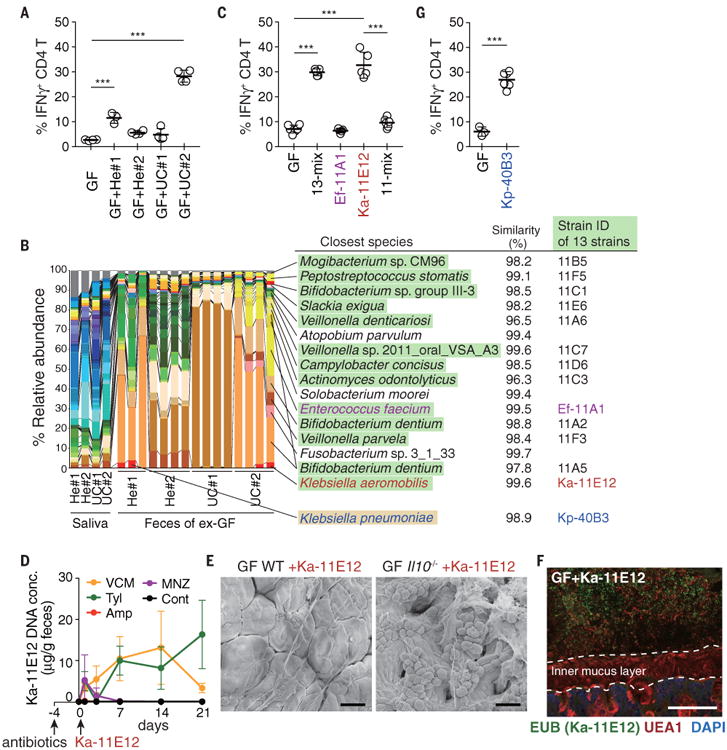
Fig. 3.

Klebsiellaaeromobilis is another TH1cell–inducing oral cavity-derived bacterial species. (A) Frequencies of IFNγ+ within colonic LP CD4+TCRβ+ T cells from exGF mice inoculated with saliva samples from healthy donors and UC patients. (B) Pyrosequencing of 16S rRNA genes of the saliva microbiota of healthy controls and patients with UC and of the resulting fecal microbiota of exGF mice (n = 3 to 4 mice per group). The relative abundance of OTUs and closest known species for each OTU are shown. OTUs corresponding to the isolated 13 strains and Kp-40B3 are marked in green and yellow, respectively. (C) The percentage of TH1 cells in the colonic LP of B6 mice colonized with 13-mix, Ef-11A1, Ka-11E12, or 11-mix. (D) SPF B6 mice were untreated or continuously treated with antibiotics in the drinking water starting 4 days before oral administration of 2 × 10⁸ CFU of Ka-11E12. Ka-11E12 abundance in fecal samples was determined by qPCR. (E) Representative SEM images of the colon of Ka-11E12–monocolonized WT or Il10-/- mice. (F) Staining with DAPI (blue), EUB338 FISH probe (green), and UEA1 (red) of the colon of mice monocolonized with Ka-11E12. Scale bars, 30 μm (E) and 100 μm (F). (G) The percentage of TH1 cells in the colonic LP of B6 mice colonized with Kp-40B3. Each point in (A), (C), and (G) represents an individual mouse (thick bars, means); points in (D) represent means of a group of five or six mice. Error bars, SD. ***P < 0.001; one-way ANOVA with post hoc Tukey's test [(A) and (C)] and two-tailed unpaired Student's t test (G).
