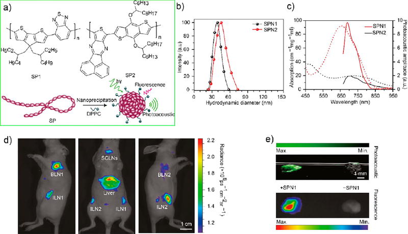
Figure 8.
(a) Molecular structures of semiconducting polymers (SP) SP1 and SP2 used for the preparation of CPNs for fluorescence and photoacoustic imaging. Schematic of the preparation of SP nanoparticles (SPNs) through nanoprecipitation. SP is represented as a long chain of chromophore units (red oval beads). 1,2-Dipalmitoyl-sn-glycero-3-phosphocholine (DPPC) is represented by a short hydrophobic tail and a charged head. (b) Representative DLS profiles of SPNs. (c) Ultraviolet–visible absorption (dashed lines) and photoacoustic spectra (solid lines) of SPNs. (d) Fluorescence/bright-field images of the corresponding mouse. (e) Ex vivo photoacoustic/ultrasound coregistered (top) and fluorescent/bright-field (bottom) images of resected lymph nodes from the mouse with SPN1 injection (50mg/mouse) (left) or a control mouse without SPN1 injection (right) in an agar phantom. Reproduced in part from Pu, K.; Shuhendler, A. J.; Jokerst, J. V.; Mei, J.; Gambhir, S. S.; Bao, Z.; Rao, J., Semiconducting polymer nanoparticles as photoacoustic molecular imaging probes in living mice. Nat. Nanotechnol. 2014, 9 (3), 233–239 (ref 63), Copyright 2014, Rights Managed by Nature Publishing Group.

