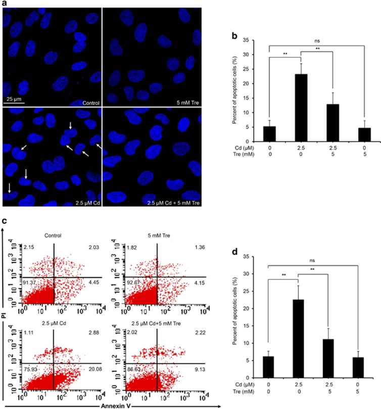
Figure 3.
Effect of Tre on Cd-induced apoptosis in rPT cells. (a and b) Cells grown on coverslips were coincubated with 2.5 μM Cd and 5 mM Tre for 12 h, and nuclear chromatin changes (apoptosis) were assessed by DAPI staining. Changes of nuclei fragmentation with condensed chromatin are evident (thin arrows). Representative morphological changes of apoptosis are present in (a), and its statistical result of apoptotic rates (b) are expressed as mean±S.E.M. (n=9). (c and d) Cells were treated with Cd and/or Tre for 12 h to assess the apoptosis using flow cytometry. Representative dot plots of Annexin V-PI staining are present in (c), and its statistical result of apoptotic rates (d) are expressed as mean±S.E.M. (n=9). NS, not significant; **P<0.01

