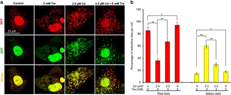
Figure 7.
Effects of Cd and/or Tre on autophagic flux in rPT cells. Cells grown on coverslips were transfected with RFP-GFP-LC3 plasmid for 36 h, and then treated with 2.5 μM Cd and/or 5 mM Tre for 12 h. (a) Representative confocal images of different treatments as indicated. (b) The number of yellow puncta (autophagosomes) and the number of red puncta (autolysosomes) in the merged images were counted and the total number of puncta per cell was calculated as percentage. Data are presented as mean±S.E.M., n=3 independent experiments; *P<0.05; **P<0.01

