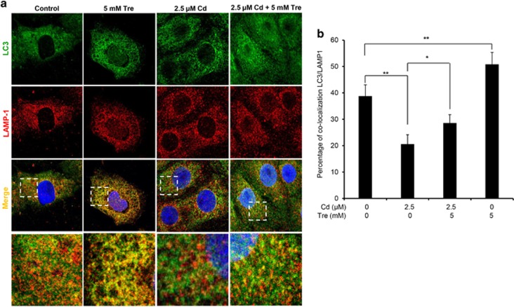
Figure 9.
Tre restored Cd-inhibited autophagosome–lysosome fusion in rPT cells. Cells grown on coverslips were treated with 2.5 μM Cd and/or 5 mM Tre for 12 h, and then successively stained with LC3 (green), LAMP-1 (red) and DAPI (blue). Colocalization of LC3 and LAMP-1 was assessed by confocal microscopy. (A) Representative confocal images showing colocalization of LC3 with LAMP-1. Higher magnification images of the outlined area are shown on the bottom. (b) Percent of colocalization of LC3 with LAMP-1. Data are represented as mean±S.E.M. of three independent experiments with 50 cells per condition in each experiment. *P<0.05; **P<0.01

