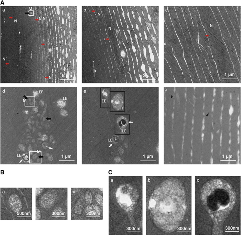
Figure 3.
Interrupted organelle degradation in the lens fiber cells of hsf4null zebrafish. (A) Transmission electron micrographs of lens fiber cells from 5-M-old hsf4null zebrafish and WT zebrafish. Nucleus (red arrow), mitochondria (black arrow) and vesicular structures (white arrow) are obvious features in hsf4null lens fiber cells (a–e). In addition, the abbreviations including N (nucleus), M (mitochondria), EE (early endosome), LE (late endosome) and L (lysosome) were marked in the picture to emphasize the organelles' persistence. The connection between the fiber cells is loose in the cortical fiber region (a and b). (c) The fiber cells in the inner region of lens have an intact nucleus. (d and e) The mitochondria and vesicular structures including EE, matured LE and LE/lysosome fusion structure depositing in the lens fiber cells. The white arrows indicate the LE/lysosome fusion structure. (f) Showing that no cell apparatus existed in the WT lens fiber cells. (B) Enlargement pictures of the white box labeling areas showing that intact mitochondria existed in lens fiber cells. Subpanel a corresponds to white box in Aa. (b and c) Upper and lower white boxes in Ad, respectively. (C) Enlargement pictures of black box labeling areas in Ae showing the endosomes at different stages. (a) EE, (b) LE and (c) LE/lysosome fusion structure

