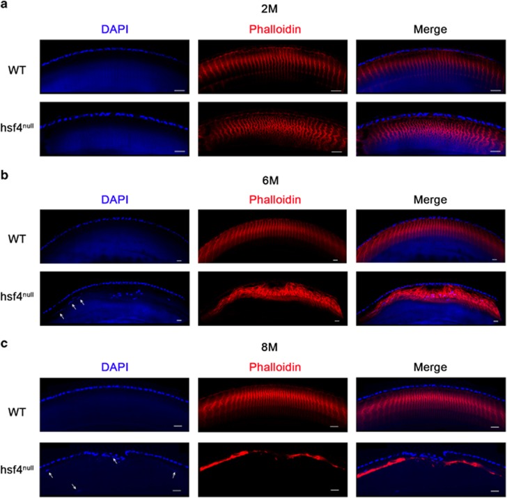
Figure 4.
Progressively disordered arrangement of lens fiber cells in the in the hsf4null zebrafish lens. Immunostaining the WT and hsf4null zebrafish lens from different ages with antiphalloidin antibody. (a) The signals are more denser in the hsf4null zebrafish than the WT zebrafish at 2M of age. (b) The fiber cell structures becoming more disorganized in the hsf4null zebrafish at 6M of age. (c) The irregular signal disappeared in the hsf4null zebrafish at 8M of age. The white arrows indicate the mislocated fiber cell nucleus in the anterior region of the lens. Scale bar: 20 μm

