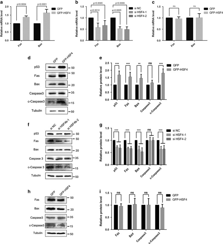
Figure 6.
HSF4 can initiate the Fas-mediated apoptosis signaling pathway in HLECs. (a) Fas and Bax were upregulated at the mRNA level when HSF4 was overexpressed in HLECs. GFP-tagged HSF4 and negative control GFP plasmids were transfected into HLE cells. And then cells were harvested for RNA extraction and real-time PCR detection. Five independent experiments were performed and the relative mRNA levels were normalized to β-actin. (b) Fas and Bas were downregulated at the mRNA level when HSF4 was silenced in HLECs. The HSF4-specific siRNAs were transfected into HLECs. And then cells were harvested for RNA extraction and real-time PCR detection. Five independent experiments were performed and the relative mRNA levels were normalized to β-actin. (c) The upregulation of FAS and BAX was p53-dependent. GFP-tagged HSF4 and negative control GFP plasmids were transfected into H1299 cells. And then cells were harvested for RNA extraction and real-time PCR detection. Five independent experiments were performed and the relative mRNA levels were normalized to β-actin. (d) HLECs were transfected with GFP-HSF4 plasmids and negative control GFP vectors. Cells were harvested 48 h after transduction followed by protein extraction and western blot detection. (e) Statistical analysis of western blot detection result in d. The relative protein level was normalized by α-tubulin. Three independent experiments were performed. (f) The HSF4-specific siRNAs were transfected into HLECs. And then cells were harvested for protein extraction and western blot detection. (g) Statistical analysis of western blot detection result in f. The relative protein level was normalized by α-tubulin. Three independent experiments were performed. (h) H1299 cells were transfected with GFP-HSF4 and negative control GFP vectors. After 48 h later, cells were harvested for protein extraction and western blot detection. (i) Statistical analysis of western blot detection result in f. The relative protein level was normalized by the α-tubulin. Three independent experiments were performed. The overexpression and RNA-interfering efficiency were validated simultaneously, and the results were listed in Supplementary Figure 5 and Supplementary Figure 6

