Figure 5.
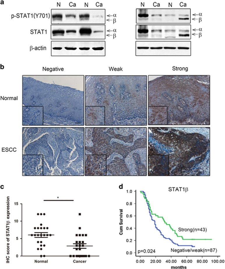
Expression of STAT1β in ESCC patient samples. (a). STAT1β expression in ESCC tumors was examined by western blot. Compared to benign esophageal tissue harvested at the surgical margins in the same specimens (labeled N), cancerous tissue (labeled Ca) often expressed a lower level of STAT1β (e.g. cases 1–3). A small subset of tumors (e.g. case 4) had high levels of STAT1β. (b) Immunohistochemistry of formalin-fixed, paraffin-embedded tissues showed variable levels of predominantly cytoplasmic STAT1β were detectable in esophageal epithelial and ESCC tissues. Based on the staining intensity, normal epithelia and tumors in our cohort were categorized into STAT1-negative, STAT1-weak or STAT1-strong (IHC stain, scale bar, 20 μm). (c) The immunohistochemistry scores of STAT1β showed that expression of STAT1β is higher in normal tissues compared to cancer tissues. (d) By Kaplan–Meier analysis, a significant correlation between overall survival and the expression level of STAT1β was found between patients with STAT1β-strong and STAT1β-weak/negative staining (*P<0.05)

