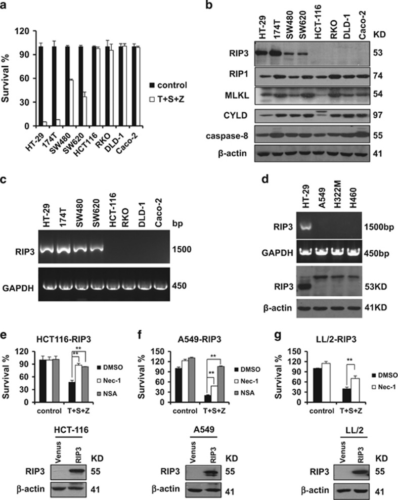
Figure 1.
The expression of RIP3 determines the sensitivity of cancer cells to necroptosis. (a) The indicated colon cancer cells were treated with DMSO (control) or TNFα(T)/Smac mimetic(S)/z-VAD(Z) for 48 h. Cell viability was determined by measuring ATP levels according to the manufacturer’s protocol (CellTiter-Glo Luminescent Cell Viability Assay; Promega). The data are represented as the mean±S.D. of duplicate wells. (b and d) Western blotting analysis of lysates from the indicated cancer cell lines to measure the protein levels of RIP1, RIP3, MLKL, CYLD, caspase-8 and β-actin. (c and d) Reverse transcription-PCR products from the indicated cells to detect the Rip3 mRNA. (e–g) The generated cancer cell lines stably expressing flag-tagged RIP3 were treated with DMSO or T/S/Z plus Nec-1 or NSA for 48 h. Cell viability was determined by measuring ATP levels. The data are represented as the mean±S.D. of duplicate wells. Abbreviations: Nec-1, Necrostatin-1; NSA, Necrosulfonamide

