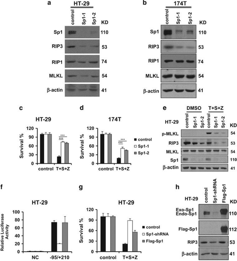
Figure 3.
Sp1 regulates the expression of RIP3 and necroptosis. (a and b) HT-29 or 174T cells stably expressing Sp1-shRNA or control vector were generated as described in the Materials and Methods. Western blotting analysis of lysates from the indicated stable cell lines showing Sp1, RIP1, RIP3, MLKL and β-actin levels. (c and d) The indicated control or Sp1-shRNA stable cell lines were treated with DMSO or T/S/Z for 24 h. Cell viability was determined by measuring ATP levels. ***P<0.001. (e) The indicated control or Sp1-shRNA stable cell lines were treated with DMSO or T/S/Z for 8 h. Cell lysates were collected and aliquots of 40 μg were subjected to western blot analysis of p-MLKL, RIP3, MLKL, Sp1 and β-actin levels. (f) Analysis for transcription activity of Rip3 promoter by the co-transfection of luciferase reporter plasmid in HT-29 cells, Sp1-shRNA and WT-Sp1 cell lines. Sp1-shRNA: HT-29 cells stably expressing an shRNA targeting Sp1. Flag-Sp1: Sp1-shRNA cells stably expressing an shRNA-resistant Sp1. (g) HT-29 cells, Sp1-shRNA and Flag-Sp1 cell lines were treated as indicated for 24 h. Cell viability was determined by measuring ATP levels. (h) Western blotting analysis of lysates from the indicated cell lines showing Sp1, RIP3 and β-actin levels

