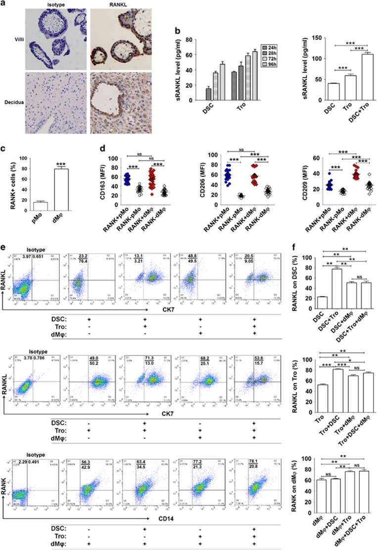
Figure 1.
The crosstalk between fetus and mothers leads to high levels of RANKL/RANK expression at the maternal–fetal interface. (a) RANKL expression in villi and decidua of normal pregnancy (n=12) by immunohistochemistry. Original magnification: × 200. (b) RANKL secretion by primary trophoblasts (1 × 10⁵ cells per well) and DSCs (1 × 10⁵ cells per well) (n=6) from normal pregnant women by ELISA after culture for 24–96 h (left). RANKL production by trophoblasts alone, DSCs alone and the co-culture of trophoblasts and DSCs (n=6) for 48 h (right). (One-way ANOVA). (c) We isolated primary PBMCs from peripheral blood (n=6) and DLC from deciduas of normal pregnant women (n=6), and then analyzed RANK expression on pMo and dMφ from normal pregnant women by labeling with anti-CD14, RANK and CD45 antibodies. (Student’s t-test). (d) Further analysis of the phenotype of RANK+ and RANK− pMo and dMφ from normal pregnant women (n=24) by FCM. (One-way ANOVA). (e and f) Trophoblasts, DSCs and/or dMφ (n=6) were co-cultured at a 1 : 1 : 1 ratio for 48 h, and then RANKL expression on CK7+ trophoblasts and CK7− DSCs, and RANK on CD14+ dMφ were evaluated by FCM, respectively. (One-way ANOVA). Tro: human trophoblasts. pMo: human peripheral blood monocytes; dMφ: human dMφ. Data are expressed as the mean±S.E.M. *P<0.05, **P<0.01 and ***P<0.001. NS: no statistical difference

