Figure 6.
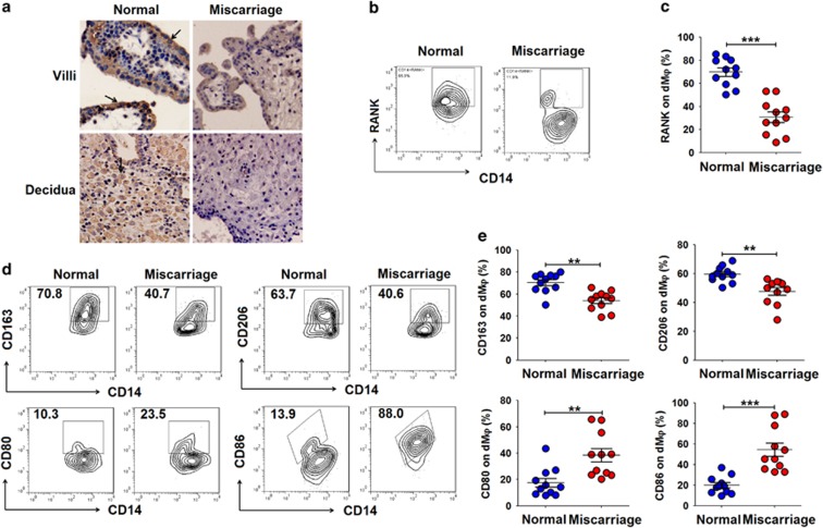
There are low levels of RANKL/RANK at the maternal–fetal interface during miscarriage. (a) Immunohistochemistry analysis of RANKL expression in villi and deciduas from women with normal pregnancy (n=12) or miscarriage (n=12) during the first trimester. RANKL expression was localized to the cell membrane and the cytoplasm (arrows) in the deciduas and villi. Original magnification: × 200. (b and c) FCM analysis of the percentage of RANK+ dMφ from women with normal pregnancy (n=11) or miscarriage (n=11) during the first-trimester. (d and e) FCM analysis of the percentage of CD163+, CD206+, CD80+ and CD86+dMφ from women with normal pregnancy (n=11) or miscarriage (n=11) during the first trimester. Normal: normal pregnant women; Miscarriage: SA women. Data are expressed as the mean±S.E.M. **P<0.01 and ***P<0.001 (Student’s t-test)

