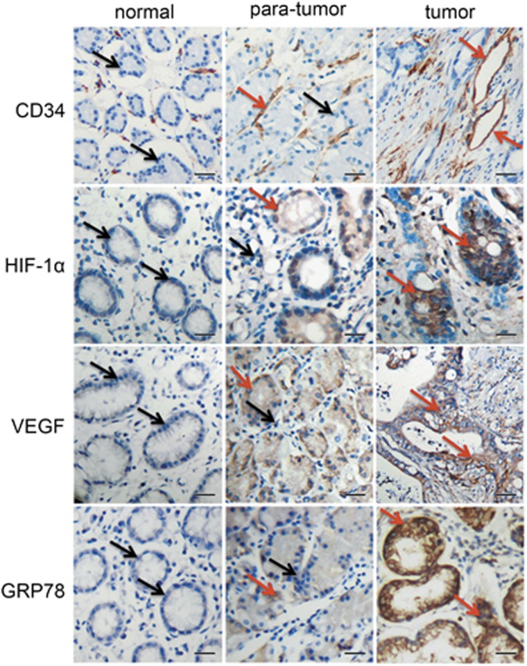
Figure 1.
Expression of CD34, HIF-1α, VEGF, and GRP78 in normal human gastric tissues, para-tumour tissues, and tumour tissues. Immunohistochemical staining of paraffin-embedded serial tissue sections from 30 normal gastric tissue specimens, 17 gastric para-tumour specimens, and 42 gastric carcinoma specimens, with antibodies against CD34, HIF-1α, VEGF, and GRP78. Immunoreactivity is indicated by brown staining. The red arrow represent positive, and the black arrow represent negative. Scale bars represent 20 μm

