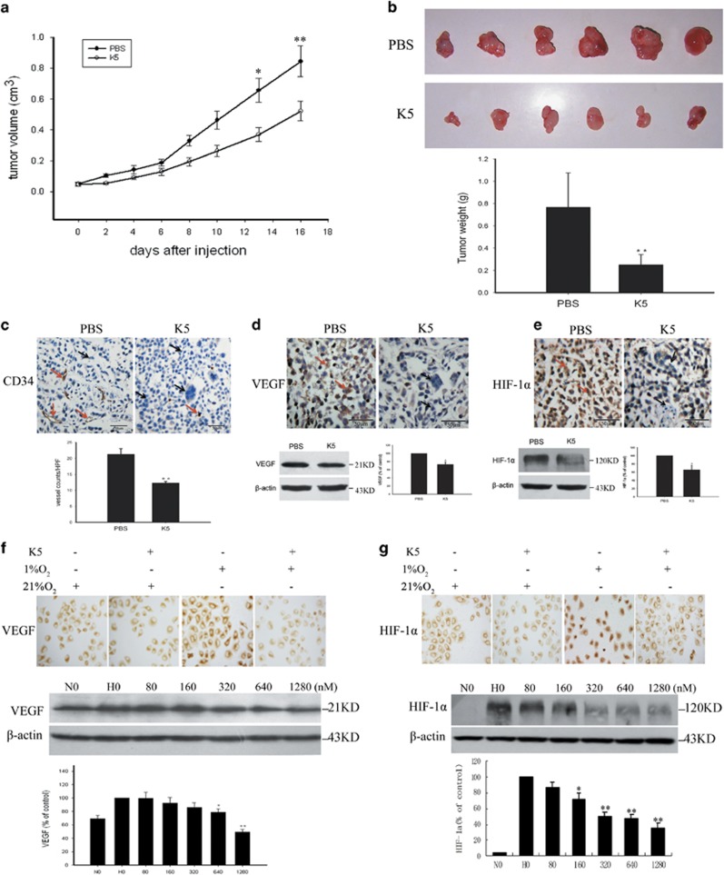
Figure 2.
K5 suppresses human gastric cancer growth and downregulates HIF-1α and VEGF. (a) Kinetics of tumour growth. Data are presented as mean±S.E.M., *P 0.05, **P<0.01. (b) Tumours were collected and weighed on day 22 after transplantation. An average suppression of 67% of primary tumour growth was observed in the K5-treated group versus PBS-treated group. **P<0.01. (c) Microvessel density was assessed by immunohistochemical staining for CD34, a marker of endothelial cells. Number of microvessels was counted from five randomly selected fields. **P<0.01. (d and e) Immunohistochemistry and western blotting were used to detect the expression of VEGF (d) and HIF-1α (e) in mice tumour tissues. Corresponding quantifications by western blotting are presented as mean±S.E.M.; n=3, *P<0.05. Scale bars represent 50 μm. (f and g) SGC-7901 cells were treated with K5 (640 nM for immunocytochemistry and 0–1280 nM for western blotting) under normoxia (21% O2) or hypoxic (1% O2) conditions for 12 h. The expression of VEGF (f) and HIF-1α (g) was determined by immunocytochemistry and western blotting analysis. Corresponding semiquantification values measured by western blotting densitometry are presented as mean±S.E.M., n=3, *P<0.05, **P<0.01. N, normoxia; H, hypoxia

