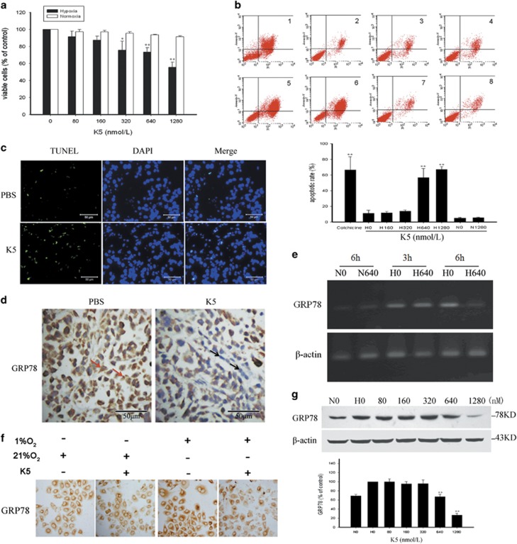
Figure 4.
K5 inhibits the proliferation of SCG-7901 cells and suppresses the expression of GRP78. (a) SGC-7901 cells were treated with the indicated concentrations of K5 for 72 h under normoxia (□) or hypoxia (▪). The viable cells were quantified by MTT. (b) SGC-7901 cells were treated with K5 (0, 160, 320, 640, or 1280 nM) for 48 h. Cells treated with 25 μM colchicine served as positive control. Apoptotic cells were quantified by flow cytometry. Data are shown as mean±S.E.M.; n=3, **P<0.01. (c) Frozen tumour sections were subjected to TUNEL assay (green), and the nuclei were counterstained with DAPI (blue) to identify apoptotic cells. Scale bars represent 50 μm. (d) Immunohistochemistry was used to detect GRP78 expression in mice tumour tissues. Scale bars represent 50 μm. (e) SGC-7901 cells were cultured with or without 640 nM K5 under normoxia or hypoxia for 3 or 6 h. GRP78 mRNA levels were analysed by reverse transcriptase-PCR. (f and g) K5 treated SGC-7901 cells were exposed to 1% or 21% O2 for 15 h and GRP 78 expression was analysed by immunocytochemistry (f) and western blotting (g). The corresponding semiquantifications by western blotting densitometry are presented as mean±S.E.M., n=3; **P<0.01. N, normoxia; H, hypoxia

