Figure 1.

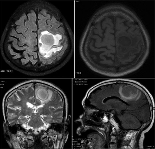
Focal rounded lesion in the cerebral parenchyma measuring 3.2 cm × 2.8 cm in the left high frontoparietal lobe. The lesion shows hypointense signal on T1 and hyperintensive signal on T2 weighted images with thin wall

Focal rounded lesion in the cerebral parenchyma measuring 3.2 cm × 2.8 cm in the left high frontoparietal lobe. The lesion shows hypointense signal on T1 and hyperintensive signal on T2 weighted images with thin wall