
Figure 2.
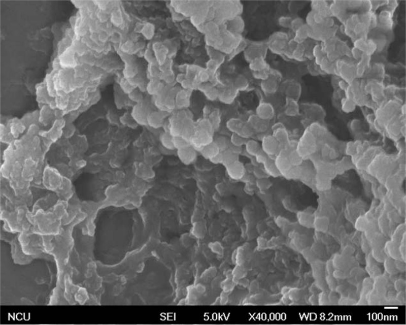
Verification of the exosomal preparation. The morphologic characterization of exosomes purified from PEs was observed by SEM, which showed round structures with heterogeneous size. PE = pleural effusion, SEM = scanning electron microscopy.
Official websites use .gov
A
.gov website belongs to an official
government organization in the United States.
Secure .gov websites use HTTPS
A lock (
) or https:// means you've safely
connected to the .gov website. Share sensitive
information only on official, secure websites.
Verification of the exosomal preparation. The morphologic characterization of exosomes purified from PEs was observed by SEM, which showed round structures with heterogeneous size. PE = pleural effusion, SEM = scanning electron microscopy.